You are viewing 1 of your 1 free articles. For unlimited access take a risk-free trial
Although uncommon, scapular fractures can seriously impact athletes who are unfortunate enough to sustain one. SPB explains how they can occur, their diagnosis, and how these injuries are rehabilitated
The scapula bone (more commonly known as the shoulder blade) is a thin, fairly flat, and roughly triangular-shaped bone, which is situated on either side of the upper back, sitting next to the back of the upper ribcage (see figure 1). This bone, along with the clavicle (collar bone) and the sternum (chest bone), constitutes what is known as the ‘pectoral-shoulder’ girdle, which connects and anchors the upper limbs to the trunk.
In the general public, most scapular fractures almost always occur as a result of a high energy-impacts, and in particular as a result of injuries sustained in motor vehicle collisions(1). Even so, scapular fractures are comparatively rare, representing just 1% of all fracture injuries(2). That said, according to the US National Trauma Data Bank, the rate of this specific injury doubled from 1% to 2.2% within a single decade, suggesting that it may be on the rise(3). Having said that, the greater use of CT scans to evaluate trauma patients (which can result in a higher detection of scapular fractures(4)) may partly help to explain this rise.
Regardless, scapular fractures are still fairly uncommon, and this is no doubt explained by the relatively unexposed, deep location of the scapula bone and the surrounding musculature. When it comes to sport, scapular fracture of the body (ie of the main portion of the bone rather than the ‘processes’ – see figure 2) is very rare. In one study, that accumulated data for 7920 player participation hours across a range of sports and 306 players, the reported rate of fractures of the scapula ranged from 1 to 10 injuries per ten thousand hours of sports participation(5).
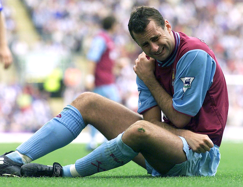
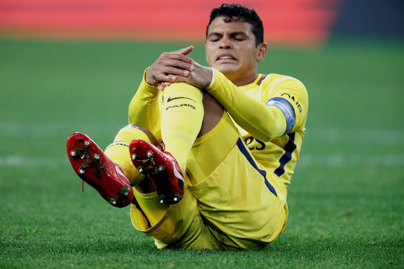
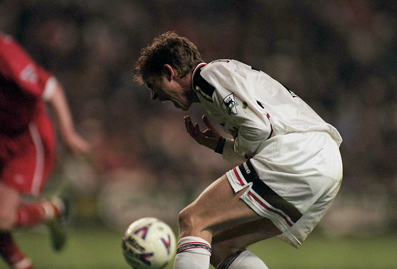
As might be expected, players in sports that involve collisions – either accidental or intentional – such as rugby and soccer are at increased risk of scapular fracture. Elite rugby players may be especially at risk as they are frequently subjected to high-energy collisions. During a tackle or collision, a player is typically subjected to impact forces in excess of double their bodyweight in the region of 400+lbs(6). Cyclists – particularly mountain bikers – may also be especially at risk as a result of falls from the bike or impact with other objects(7).
In one review study into scapular body fracture in sport, researchers determined that acute trauma to the shoulder was responsible for 70% of injuries(8). However, these injuries occurred as a result of both contact and non-contact mechanisms. Athletes participating in American football, wrestling, and water skiing tended to suffer injury as a result of forceful rotation movements, while injuries as a result of forceful swinging movements were recorded in ice hockey, boxing, and badminton. Fatigue fractures were rarer still; mechanisms included repetitive stress from pitching in a baseball player, an extended season of batting in a cricket player, and jogging with hand weights in a runner.
As figure 1 shows, the scapular is a large structure with various regions and bony protrusions, which can experience different loading forces depending on the movement of the arm. This in turn means that different types of scapular fracture can occur at different locations (as shown in figure 2). In addition to location, researchers have produced a system of scapula fracture classification based on the mechanism of injury (see figure 3)(6). These are as follows:
· *Type A: direct impact fracture involving the scapula neck and body.
· *Type B: fracture affecting the articular surface caused by an outstretched arm.
· *Type C: ‘twisting’ type of fracture, which extends into the region of the scapula known as the ‘coracoids’.
As mentioned above, while the majority of scapular fractures in athletes are due to direct impacts or excessive forces sustained during falls or collisions, a fatigue fracture of the scapular body – while rarer still – is occasionally observed. Unlike other scapular body fractures (which also occur in the general population), fatigue fractures have only ever been documented in athletes(9). The mechanism of a scapular body fatigue fracture appears to involve repetitive trauma across the bone (as a result of cumulative muscular contractions). This results in micro-fractures occurring in the scapular body(10). The more subtle nature of the fracture in this injury mechanism/type makes a diagnosis much more difficult, even when high resolution imaging is used such as MRI scanning (see later).
With few cases of sports-related scapular fracture in the medical literature, there is no universally-agreed protocol for diagnosis of this type of injury. This is also complicated by the fact that the types of symptoms experienced following a scapula fracture are also dependent on the exact location of the fracture. However, some of the commonly reported symptoms can typically be described as follows(11):
· In most cases, a sudden onset of pain occurs, usually localized across the shoulder blade and/or at the top of the shoulder – but which may also be diffuse - and which worsens with time. (NB: note too that, although extremely rare, a fatigue fracture of the scapula will likely present with a much more gradual pain onset.
· Pain that is aggravated by arm movement or deep inhalation.
· Pain that is severe when the patient attempts to raise the arm overhead.
· Range of motion that is severely limited, particularly with abduction (moving the arm outwards from the body).
· An absence of ‘popping’ sensations during movement (popping sensations during movement generally indicate cartilage damage) but possibly accompanied instead by a ‘grinding’ sensation.
· Swelling, tenderness and bruising in the top part of scapula region, or on the top of the shoulder towards the end of the collarbone (clavicle).
· When viewed from the injured side, the shoulder girdle may take on a slightly flattened or deformed appearance compared to the unaffected side.
In addition to the symptoms present, the physio/clinician treating the athlete should make sure they gather a detailed injury history – particularly of any previous shoulder injuries. That’s because previous injury of the scapular is itself known to be a significant risk factor for future injury(9). If a scapular fracture is suspected, the first port of call will be an X-ray for the area. Often, this is enough for a physician or physio to make a definite diagnosis – not just of the presence of a scapular fracture, but the fracture type too.
However, according to the injury mechanism system described in figure 3 above, type B and C fractures are not always identifiable using X-ray imaging alone. Therefore, if an athlete’s injury was known to involve either an outstretched arm or a combination of abduction and external rotation therefore CT or MRI scans may also be required. Likewise, if the athlete’s physiotherapist suspects a fatigue fracture, the micro-cracks in the bone will likely not be visible on a standard X-ray, even when the athlete’s symptoms have been present for several weeks or months. In this case, MRI or CT imaging should be the first port of call rather than X-ray imaging.
Unlike other fracture injuries, there are no universally agreed rehab protocols for returning athletes to sport(12). However, the nature and mechanism of the injury are thought to provide useful guidelines as to the best treatment options. For many scapular body fractures, a conservative treatment approach without the use of surgical intervention is likely to be successful(13). One reason for this is that the vast majority of scapular body fractures involve only minimal displacement (ie minimal separation of bone segments) – thanks to the thick, strong support provided by the surrounding muscle mass. This musculature also explains why surgical repair should be considered only as a last resort; surgery will often requires a fair amount of cutting of muscles, which results in a much a longer recovery.
The typical rehab protocol for conservative treatment is as follows (timeframe 2-6 months(6)):
· Immobilization using a sling and swathe for comfort (for up to six weeks).
· As soon as pain begins to subside, begin progressive range of motion exercises.
· Once pain free, begin progressive loading against resistance (concentrating on developing maximum range of motion) – for example using Therabands.
· Assuming movements are pain free and follow up imaging confirms fracture healing (ie bone union) is taking place, begin introducing heavier and more sports-specific loading.
· Return to sport can be considered once full range of motion and bilateral strength symmetry has been achieved, and no pain is present.
Even with widely displaced scapular body fractures (ie where the fracture gap is significant), this treatment approach is likely to produce reliable fracture healing with restored shoulder function(14).
Sometimes, the type and nature of a scapular fracture means that surgical intervention is recommended, as is the case where conventional rehab protocols such as those described above have failed. In recent years, key criteria suggesting that surgery is required have been identified(15,16). It’s beyond the scope of this article to discuss these criteria here; however, they are most likely to be in those fractures sustained via an outstretched arm (type B) and those resulting from abduction plus external rotation (type C)(6). This underlines the importance of confirming (if possible) the injury mechanism.
The surgery required will involve surgical fixation (pinning the displace segments of bone back together), followed by an extended recovery period to allow bone healing and reunion to take place. Once this is deemed to be satisfactory by the surgeon, conventional rehab and physiotherapy can begin. The good news for athletes however is that while the recovery process is much longer, the outcomes following a surgical scapular fracture repair are typically equally as good as those not requiring surgery!
1. J Trauma. 1980;20(10):880-883.
2. J Am Acad Orthop Surg. 1995;3(1):22-33
3. National Trauma Data Bank. Injury. 2019;50(2):376–381
4. Era. Acad Emerg Med. 2018;25(7):738–743
5. J Sci Med Sport. 2015;18(5):529–33
6. Orthop J Sports Med. 2019 Dec 2;7(12):2325967119887388
7. BMJ Open Sport Exerc Med. 2016 Jan 11;2(1):e000042
8. Curr Rev Musculoskelet Med. 2019 Mar; 12(1): 13–23
9. HSSJ (2018) 14:328–332
10. Skelet Radiol. 1982;9(1):27–32
11. Sports Health. 2012 Nov; 4(6): 502–503
12. J Bone Joint Surg Br Vol. 1984;66(5):725–31
13. Clin Orthop Relat Res. 1991;(269):174–180
14. Operative Orthopaedics. Philadelphia: JB Lippincott; 1988:197–202
15 Clin Orthop Surg. 2023 Oct; 15(5): 695–703
16. EFORT Open Rev. 2021 Jun; 6(6): 518–525
Today you have the chance to join a group of athletes, and sports coaches/trainers who all have something special in common...
They use the latest research to improve performance for themselves and their clients - both athletes and sports teams - with help from global specialists in the fields of sports science, sports medicine and sports psychology.
They do this by reading Sports Performance Bulletin, an easy-to-digest but serious-minded journal dedicated to high performance sports. SPB offers a wealth of information and insight into the latest research, in an easily-accessible and understood format, along with a wealth of practical recommendations.
*includes 3 coaching manuals
Get Inspired
All the latest techniques and approaches
Sports Performance Bulletin helps dedicated endurance athletes improve their performance. Sense-checking the latest sports science research, and sourcing evidence and case studies to support findings, Sports Performance Bulletin turns proven insights into easily digestible practical advice. Supporting athletes, coaches and professionals who wish to ensure their guidance and programmes are kept right up to date and based on credible science.