You are viewing 1 of your 1 free articles. For unlimited access take a risk-free trial
Slipping rib syndrome: a pain in the upper back
Andrew Hamilton investigates a potentially painful condition in athletes known as ‘slipping rib syndrome’. What causes it, how is it treated and what should athletes, coaches and physios be on the lookout for?
Although there’s an abundance of research on lumbar (back) and cervical (neck) pain and dysfunction, there’s a relative paucity of research relating to the thoracic (mid and upper back) region(1) This is partly due to the overall rigidity of the spine as it passes through the thoracic structure, which reduces the likelihood of injury compared to freely-moving joints undergoing a variety of loading forces. However, while the thoracic structures lack the mobility of many other joint systems in the human body, some movement is required, bringing into play a number of rib joints known as ‘costovertebral’ joints and their associated ligaments that hold them in place.
The thoracic region can move!
The thorax is comprised of a number of rigid structures, including the ribs, sternum (chest bone) and thoracic vertebrae of the spine. However, the intervertebral discs of the spine and the costal (rib joint) cartilages do enable some joint movement in the thoracic region. This movement includes(2):
· Flexion-extension (forwards/backwards motion) – ranging from 4o at T1 (just below the neck) to 12o degrees at T12 (just above the lumbar region).
· Lateral (side-to-side) movement – this typically allows 6-7o per vertebral segment.
· Rotation – ranging from 9o at T1 to 2o at T12.
Although each individual vertebra joint motion is small, the overall result is that the costovertebral joints and ligaments combine to play a critical role in the movement required not only during the ribcage efforts and movement when breathing, but also in allowing thoracic mobility, while at the same time facilitating load bearing and stabilization(3).
The rib-vertebrae interaction
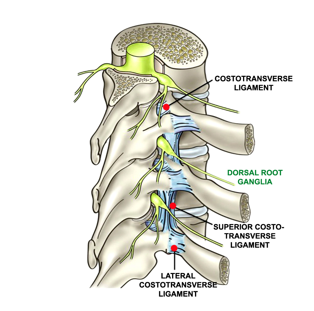
The rib-vertebra-sternum interface is critical for correct thoracic function; it needs to be rigid enough for load bearing (to protect the lungs) and stabilisation but mobile enough to allow necessary movement. Critical to this function are the costovertebral joints and their associated ligaments (see figure 1). These joints and ligaments are poorly understood and frequently overlooked anatomical structures(4), despite their abundance in the human thoracic spine (there are 108). The costovertebral ligaments are dense, fibrous ligaments, containing a large amount of elastic fibres. These fibers are able to stretch with movement in one direction yet shorten with movement in the opposite direction; this means that when subject to repeated movements, they can remain taut rather than becoming lax(5).
Figure 1: Costovertebral joint and ligament structures*
Costovertebral strain
When compression and twisting forces applied to the costovertebral joint are greater than it can withstand, a costovertebral strain can occur. This can result in damage to the costovertebral ligaments, cartilage or both. When a costovertebral strain injury occurs in an athlete, he/she will typically experience an intermittent sharp ‘stabbing pain’ followed by a dull achy sensation, which lasts for days, weeks or in severe case, even months(6,7). This pain is usually (though not always) is felt on one side (unilateral), and exacerbated by movement of the spine, especially twisting and side bending. Deep breathing, coughing and sneezing typically also produces pain in the mid or upper back, which can also radiate to the scapular (shoulder blade) region or front of the chest wall(8,9). In more severe cases, even simple activities such as rising from a chair, stretching, and turning over in bed can result in severe discomfort(10).
Diagnosing a costovertebral strain
When an athlete suffers with posterior thoracic pain (ie in the upper or mid back), correctly determining the cause of the condition can be difficult. This is partly due to the neural complexity of the thoracic spine, which is a region where many nerves pass through. Also, there are a number of other conditions that can present with similar symptoms. These include (in no particular order)(4):
· Rib/vertebra fracture.
· A thoracic intervertebral disc protrusion/herniation (where the discs bulges and impinges on a nearby nerve).
· Spinal stenosis, where the narrowing of one or more spaces within the spinal canal compresses the spinal cord and nerve roots.
· Paraspinal muscle or ligament strains (these stabilizing muscles lie close to the thoracic vertebra).
· Thoracic outlet syndrome (compression of the nerves or blood vessels in nearby tissues).
· Osteoarthritis.
· Disease of the facet joints (where the ribs articulate against the thoracic vertebrae).
· Pain resulting from other (potentially serious) conditions, including pain of cardiac, aortic, renal, pulmonary, oesopha geal or gallbladder origin, and pain from metastatic malignant lesions.
If an athlete develops thoracic pain where there is no history of impact, trauma or loading prior to the pain, further investigations are recommended to rule out more serious conditions. This is particularly true for older athletes (over the age of 45) where pain could arise from a number of disease conditions (as indicated above)(4). These investigations may include X-ray imaging, MRI and/or CT scans and ultrasound imaging. In addition to imaging, a good physiotherapist will be able to help rule conditions in or out by a process of differential diagnosis using mobility/pain tests, the patient’s history and the presence or absence of other symptoms.
Mysterious pain onset: slipping rib?
When athletes develop posterior thoracic pain, there is usually a clear onset trigger – for example an unusual loading or impact event, or the start of a new and unaccustomed mode of training. However, this is not always the case, even where further testing described above shows no other condition/disease is present. This can be very frustrating; when other injuries or conditions have been ruled out, athletes and their physios may feel they have drawn a complete blank. However, there exists another possible cause, which although uncommon, is frequently misdiagnosed or underdiagnosed – so-called ‘slipping rib syndrome’.
Slipping rib syndrome (abbreviated SRS) is a condition that can arise from the hypermobility of the front ends of the false rib costal cartilages (see figure 2). Also referred to by a number of other terms such as ‘clicking rib’, ‘interchondral subluxation’, ‘painful rib syndrome’, and ‘traumatic intercostals neuritis’, undiagnosed slipping rib syndrome can subsequently lead to months or even years of unresolved thoracic pain(11). This syndrome was first described in 1919, but while there have been many scientific papers on this condition, it is rarely mentioned in medical textbooks(12).
The primary cause of SRS is rib hypermobility, which is caused by the relative weakness of the ligaments around the costovertebral joints. In particular, the false ribs (ribs 8, 9 and 10 – lower left in figure 2) are most susceptible to hypermobility. This is because these ribs do not form a rigid continuum spanning the thoracic vertebrae and the sternum; instead of attaching to the solid and rigid sternum bone at the anterior (chest) end, they are connected to each other via a cartilaginous/fibrous band.
Figure 2: Rib structure and false rib costal cartilages

Surgical studies have shown that this hypermobility of the anterior (chest) ends of the false rib costal cartilages may lead to slipping of the affected rib under the superior adjacent rib, resulting in intercostal nerve irritation, strain of the intercostal muscles and costovertebral strain(13,14) While this false rib hypermobility stems from the anterior ends of the false rib costal cartilages, it is important to appreciate that the rib cage system is a closed one. When one end of the rib moves, the other end moves too, which means that rib hypermobility at the front (chest) end inevitably leads to problems in the posterior thoracic area (such as costovertebral strain), leading to pain (and very often) accompanied by a ‘clicking sensation’ during movement.
Homing in on SRS
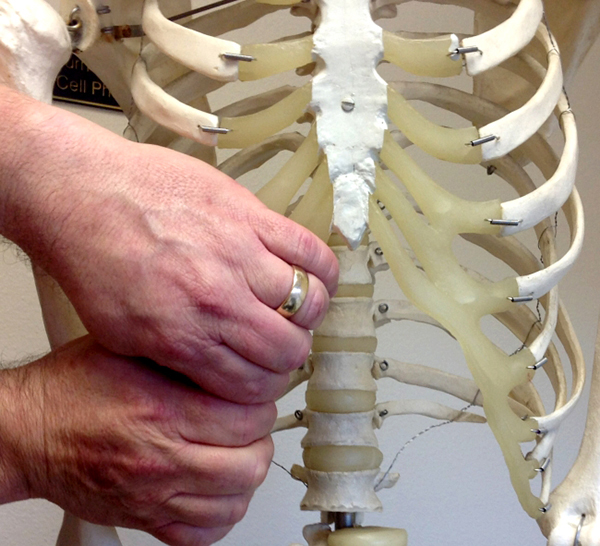
SRS can occur both as a result of an impact to the ribs and frustratingly (as we have mentioned above) spontaneously and without any prior trauma. Where SRS is the cause of thoracic pain, X-rays and and imaging tests such as MRI are unlikely to be useful(11). There is however a relatively simple clinical test that (when combined with imaging and patient history information) can help diagnose SRS. Known as the ‘hooking manoeuvre’, this test was first described by clinicians back in 1977(15). To perform this test, the physiotherapist places his or her fingers under the lower costal margin and pulls the hand in an towards the front – ie forwards direction (see figure 3). If this movement causes pain or clicking, this indicates a positive test – ie SRS is present. To confirm the diagnosis however, the use of a rib block analgesic is recommended, where a local anesthetic is applied to the intercostals joints; an absence of pain following a repeat of this test confirms a diagnosis SRS(16).
Figure 3: The ‘hooking manoeuvre’
Treatment options and prognosis
In mild cases of SRS, many athletes may experience success and complete resolution of symptoms with an initial period of rest and the avoidance of movements or postures that caused or exacerbate the symptoms. This can be followed by various conservative treatments(11), which include soft-tissue mobilisation such as massage and myofascial release, as well as other mobilisation stretching and strengthening activities. Anti-inflammatory pain relief such as Ibuprofen can also be helpful. The application of ice in the early stages has also been shown to help(17).
Research shows that many athletes with SRS that has persisted for a while may struggle psychologically due to their inability to continue to compete(17). Therefore if you suffer from this condition or coach athletes who do, you should take comfort from the fact that in the majority of cases, SRS is a benign condition that is most definitely treatable. If initial rest, ice, stretching, massage and anti-inflammatories fail to resolve SRS, other conservative treatments may help, including heat, chiropractic manipulation and electronic stimulation, all of which focus on symptom control(18). The keys goals of treatment should always be focussed on the control of inflammation and pain.
When SRS doesn’t easily resolve
When conservative treatment methods such as those outlined above are unsuccessful, minimally invasive treatment should be considered starting with a combination of steroids and local anesthetics(19). These local nerve blocks will not only provide symptomatic relief, but will also confirm the diagnosis and rule out other causes such as fracture and impact damage. In some cases, steroids and local anesthetics will largely abolish the symptoms, which may not recur for a significant period of time.
Unfortunately however, it may be that the measures above do not produce a resolution, or that they only help for a brief while. In chronic and stubborn cases of SRS, the only long-term solution is correcting the underlying rib anatomy, especially given the given the anatomical causes of the pain. In order to change the anatomy and definitively cure SRS, surgical intervention is recommended, which often involves the resectioning of any abnormal cartilaginous attachment of the affect rib(s). Although the rehabilitation period for surgery is much lengthier, the long-term outcome is still good.
In summary
Slipping rib syndrome has been described in scientific literature over the span of more than one hundred years, yet it is still frequently undiagnosed or misdiagnosed. This can leave patients in pain and untreated for considerable lengths of time, despite the availability of effective treatments that carry relative low risk. For athletes who are unable to train yet have no idea what is causing them pain, this can be incredibly soul destroying.
When rib pain strikes, seeking out a physiotherapist or clinician with experience in rib injuries is therefore definitely recommended. Not only does an SRS diagnosis open the door to effective treatments, an even more profound benefit may be that of reassurance. For an athlete who has been suffering thoracic pain for weeks or even months with no obvious cause, to hear that there is nothing seriously wrong is extremely comforting. Crucially, it is also the first step to setting them on the road to recovery!
References
1. Journal of Manual and Manipulative Therapy. 2003;11:43– 48.
2. Comput Methods Biomech Biomed Engin. 2015;18:556–570
3. Cureus. 2016 Nov; 8(11): e874
4. Phys Ther. 2006;86:254–268
5. Hollinshead’s Textbook of Anatomy. New York Press 1974
6. Br J Surg. 1984;71:522–523
7. Geriatrics. 1995;50:46–49
8. Topics in Clinical Chiropractic. 1999;6:20 –38
9. Topics in Clinical Chiropractic. 1999;6:79 –92 4,10
10. Postgrad Med. 1989;86:75–78
11. Journal of Athletic Training 2005;40(2):120–122
12. Practitioner. 1919;102:314–322
13. J Pediatr Surg. 1997;32:1081–1082
14. NY State J Med. 1963;63:1670–1675
15. Am Med Assoc. 1977; 237:794–795
16. Med Sci Sports Exerc. 2003;35: 1634–1637
17. Clin J Sport Med. 2019;29(1):18–23
18. J Athl Train. 2005;40(2):120–122
19. Lancet. 2014;383(9919):844
Newsletter Sign Up
Testimonials
Dr. Alexandra Fandetti-Robin, Back & Body Chiropractic
Elspeth Cowell MSCh DpodM SRCh HCPC reg
William Hunter, Nuffield Health
Newsletter Sign Up
Coaches Testimonials
Dr. Alexandra Fandetti-Robin, Back & Body Chiropractic
Elspeth Cowell MSCh DpodM SRCh HCPC reg
William Hunter, Nuffield Health
Keep up with latest sports science research and apply it to maximize performance
Today you have the chance to join a group of athletes, and sports coaches/trainers who all have something special in common...
They use the latest research to improve performance for themselves and their clients - both athletes and sports teams - with help from global specialists in the fields of sports science, sports medicine and sports psychology.
They do this by reading Sports Performance Bulletin, an easy-to-digest but serious-minded journal dedicated to high performance sports. SPB offers a wealth of information and insight into the latest research, in an easily-accessible and understood format, along with a wealth of practical recommendations.
*includes 3 coaching manuals
Get Inspired
All the latest techniques and approaches
Sports Performance Bulletin helps dedicated endurance athletes improve their performance. Sense-checking the latest sports science research, and sourcing evidence and case studies to support findings, Sports Performance Bulletin turns proven insights into easily digestible practical advice. Supporting athletes, coaches and professionals who wish to ensure their guidance and programmes are kept right up to date and based on credible science.
